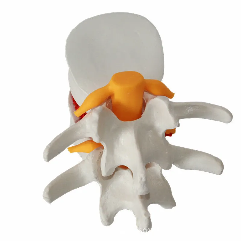
Medical teaching demonstration model of human lumbar intervertebral disc with herniation, anatomical spine model made from PVC material, measuring 11x10x7.6 cm for educational training in physiotherapy and medical schools, shows disc prolapse condition


Medical teaching demonstration model of human lumbar intervertebral disc with herniation, anatomical spine model made from PVC material, measuring 11x10x7.6 cm for educational training in physiotherapy and medical schools, shows disc prolapse condition
Medical demonstration model of lumbar intervertebral disc herniation for anatomical teaching and training

This anatomical demonstration model serves as a specialised educational tool focused specifically on lumbar intervertebral disc herniation, providing a tangible visual reference for medical and healthcare training scenarios. Designed for clarity and educational utility, the model represents a single lumbar disc in a prolapsed state, allowing trainers, physiotherapists, and medical educators to demonstrate the anatomical changes associated with this common spinal condition. Its compact dimensions make it suitable for classroom demonstrations, clinical consultations, and individual study sessions where understanding disc pathology is essential for treatment planning and patient education.
Features and Construction
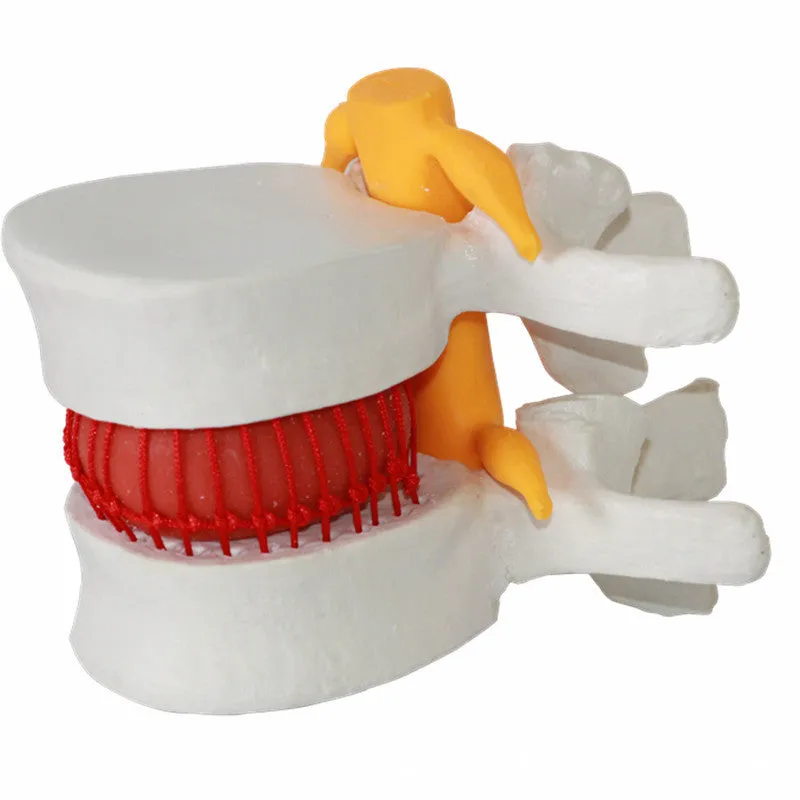
Constructed to show specific anatomical details relevant to intervertebral disc disorders, this model prioritises educational clarity over comprehensive spinal representation. The focus remains solely on the lumbar disc region, with particular attention to the herniation pathology that characterises many back pain conditions. This targeted approach makes the model particularly valuable for specialised training in orthopaedics, physiotherapy, and chiropractic education where disc pathology requires detailed explanation.
Material and Build
The model utilises PVC (polyvinyl chloride) as its primary construction material, chosen for its ability to maintain structural integrity while allowing detailed anatomical representation. This material selection provides sufficient rigidity to preserve the model's shape during repeated handling in educational environments, while the white colour finish offers clear visual contrast against typical classroom backgrounds. The material properties support the model's function as a demonstration tool rather than a fully flexible anatomical replica, prioritising clarity of the herniation depiction over biomechanical simulation.
Size and Practical Fit
With dimensions of 11 cm in length, 10 cm in width, and 7.6 cm in height, the model maintains a compact form factor suitable for tabletop demonstrations and individual examination. The 0.22 kg weight ensures easy portability between teaching locations while providing enough substance for comfortable handling during explanations. These practical dimensions allow the model to be conveniently stored in educational cabinets, transported in professional bags, or displayed on consultation room desks without occupying excessive space, making it accessible for both group teaching and one-to-one patient education scenarios.
Uses and Placement

This demonstration model addresses specific educational needs across multiple healthcare training environments, providing a visual anchor for discussions about spinal anatomy and pathology. Its specialised focus on disc herniation makes it particularly relevant for contexts where understanding this specific condition forms a core component of professional knowledge or patient communication.
Event or Professional Use
In formal educational settings, the model serves medical schools, physiotherapy training programmes, and continuing professional development workshops where spinal anatomy forms part of the curriculum. Clinical educators can use it to demonstrate the anatomical basis of disc prolapse during lectures, small group tutorials, or practical examination preparation. Healthcare professionals may incorporate it into patient consultations to visually explain diagnostic findings, surgical procedures, or conservative treatment approaches for lumbar disc disorders, bridging the gap between medical terminology and patient understanding.
Everyday Home Use
While primarily designed for professional educational contexts, the model may also support self-directed learning for healthcare students studying spinal anatomy outside formal classroom environments. Its clear representation of disc herniation can aid revision of pathological conditions for examination preparation or clinical placement reflection. The compact size allows for convenient storage on study desks or bookshelves, providing an immediate reference when reviewing notes or preparing presentations on spinal disorders without requiring access to larger anatomical collections.
Benefits and Buying Value

This specialised demonstration model offers practical advantages for targeted anatomical education, focusing resources on a specific pathological condition rather than attempting comprehensive spinal representation. The value derives from its specialised educational function and durable construction suited to repeated handling in teaching scenarios.
Reuse and Low Maintenance
The PVC construction provides material durability appropriate for repeated handling in educational environments, maintaining anatomical details through multiple teaching sessions. The model requires minimal maintenance beyond occasional dusting and careful storage in its packaging between uses. This low-maintenance approach supports extended educational utility across multiple student cohorts or patient consultation sessions, representing a sustainable investment for training programmes with recurring need for disc pathology demonstration.
Why Choose This Product
Select this model for its specific focus on lumbar intervertebral disc herniation—a common pathological condition requiring clear visual explanation in medical and physiotherapy education. The compact dimensions and manageable weight facilitate practical use in various teaching environments, from lecture theatres to consultation rooms. The white PVC construction offers clear visual definition of anatomical structures, while the packaging dimensions ensure secure storage and transport. This combination of specialised educational focus and practical design addresses a recognised need in anatomical teaching without the complexity or expense of more comprehensive spinal models.